多数人(高达60%)感染贝氏柯克斯体无症状。[2]Hartzell JD, Wood-Morris RN, Martinez LJ, et al. Q fever: Epidemiology, diagnosis, and treatment. Mayo Clin Proc. 2008;83:574-579.http://www.mayoclinicproceedings.org/article/S0025-6196(11)60733-7/fulltexthttp://www.ncbi.nlm.nih.gov/pubmed/18452690?tool=bestpractice.com[5]Maurin M, Raoult D. Q fever. Clin Microbiol Rev. 1999;12:518-553.http://cmr.asm.org/content/12/4/518.fullhttp://www.ncbi.nlm.nih.gov/pubmed/10515901?tool=bestpractice.com已有超过30个不同的临床症状被描述。然而,急性感染一般表现为流感样症状伴有不同程度的肺炎和肝炎。大多数患者症状轻微且多为自限性,一般2周内自行缓解。[5]Maurin M, Raoult D. Q fever. Clin Microbiol Rev. 1999;12:518-553.http://cmr.asm.org/content/12/4/518.fullhttp://www.ncbi.nlm.nih.gov/pubmed/10515901?tool=bestpractice.com[6]Parker NR, Barralet JH, Bell AM. Q fever. Lancet. 2006;367:679-688.http://www.ncbi.nlm.nih.gov/pubmed/16503466?tool=bestpractice.com
在一小部分患者中,原发感染可导致持续性局灶性感染(例如:心内膜炎、血管感染、骨关节感染、淋巴结炎)。[3]Eldin C, Mélenotte C, Mediannikov O, et al. From Q fever to Coxiella burnetii infection: a paradigm change. Clin Microbiol Rev. 2017;30:115-190.http://www.ncbi.nlm.nih.gov/pubmed/27856520?tool=bestpractice.com持续性局灶性感染的危险因素包括妊娠、[51]Baud D, Greub G. Intracellular bacteria and adverse pregnancy outcomes. Clin Microbiol Infect. 2011:17;1312-1322.http://www.ncbi.nlm.nih.gov/pubmed/21884294?tool=bestpractice.com之前存在心脏疾病(例如风湿热、二叶主动脉瓣、先天性心脏病、假体心脏瓣膜、瓣膜反流、狭窄≥II 度或二尖瓣脱垂病史)、癌症(例如:血液系统恶性肿瘤)、血管病变(例如血管移植、血管动脉瘤)和 HIV 感染或化疗导致的免疫功能受损状态。[5]Maurin M, Raoult D. Q fever. Clin Microbiol Rev. 1999;12:518-553.http://cmr.asm.org/content/12/4/518.fullhttp://www.ncbi.nlm.nih.gov/pubmed/10515901?tool=bestpractice.com心内膜炎是持续性局灶性感染的最常见形式(高达 70% 的病例)。[2]Hartzell JD, Wood-Morris RN, Martinez LJ, et al. Q fever: Epidemiology, diagnosis, and treatment. Mayo Clin Proc. 2008;83:574-579.http://www.mayoclinicproceedings.org/article/S0025-6196(11)60733-7/fulltexthttp://www.ncbi.nlm.nih.gov/pubmed/18452690?tool=bestpractice.com
发热患者伴有非特异性症状时需要高度怀疑贝氏柯克斯体感染的诊断。非常罕见报告近期接触临产动物。大多实验室无法培养贝氏柯克斯体,是因为存在技术困难。培养贝氏柯克斯体必须在生物安全 3 级防护,因其病原微生物具有强传染性且有可能成为生物恐怖武器。[2]Hartzell JD, Wood-Morris RN, Martinez LJ, et al. Q fever: Epidemiology, diagnosis, and treatment. Mayo Clin Proc. 2008;83:574-579.http://www.mayoclinicproceedings.org/article/S0025-6196(11)60733-7/fulltexthttp://www.ncbi.nlm.nih.gov/pubmed/18452690?tool=bestpractice.com[56]Raoult D, Marrie T, Mege J. Natural history and pathophysiology of Q fever. Lancet Inf Dis. 2005;5:219-226.http://www.ncbi.nlm.nih.gov/pubmed/15792739?tool=bestpractice.com因此,诊断依赖于血清学检测。
病史
应当仔细询问是否最近动物接触和/或居住或前往流行地区旅行,但经常缺乏暴露史。其他危险因素包括男性和年龄为 30-70 岁。临床医生应当询问之前存在的心脏疾病(例如风湿热、二叶主动脉瓣、先天性心脏病、假体心脏瓣膜、瓣膜反流、狭窄≥II 度或二尖瓣脱垂病史)、免疫抑制、血管异常或妊娠,因为这些疾病容易引发持续性局灶性感染。急性疾病后 3 个月至 17 年均可发生持续性局灶性感染,[57]Wilson HG, Neilson GH, Galea EG, et al. Q fever endocarditis in Queensland. Circulation. 1976;53:680-684.http://www.ncbi.nlm.nih.gov/pubmed/1253390?tool=bestpractice.com也可在没有急性疾病病史的情况下发生。[6]Parker NR, Barralet JH, Bell AM. Q fever. Lancet. 2006;367:679-688.http://www.ncbi.nlm.nih.gov/pubmed/16503466?tool=bestpractice.com
体格检查
下列是急性感染和持续性局灶性感染的可能表现:
急性感染[6]Parker NR, Barralet JH, Bell AM. Q fever. Lancet. 2006;367:679-688.http://www.ncbi.nlm.nih.gov/pubmed/16503466?tool=bestpractice.com[1]Marrie TJ, Raoult D. Coxiella burnetii. In Mandell GL, Bennett JE, Dolin R, eds. Principles and practice of infectious diseases. 6th ed. Philadelphia, PA: Churchill Livingstone; 2005.[2]Hartzell JD, Wood-Morris RN, Martinez LJ, et al. Q fever: Epidemiology, diagnosis, and treatment. Mayo Clin Proc. 2008;83:574-579.http://www.mayoclinicproceedings.org/article/S0025-6196(11)60733-7/fulltexthttp://www.ncbi.nlm.nih.gov/pubmed/18452690?tool=bestpractice.com[3]Eldin C, Mélenotte C, Mediannikov O, et al. From Q fever to Coxiella burnetii infection: a paradigm change. Clin Microbiol Rev. 2017;30:115-190.http://www.ncbi.nlm.nih.gov/pubmed/27856520?tool=bestpractice.com
常见表现:
单纯性发热,可能持续较长时间
流感样症状
Pneumonia
肝炎
不常见的表现:
心血管:急性心内膜炎、心肌炎、心包炎、心肌心包炎、动脉或静脉血栓形成
神经系统:脑膜脑炎、脑膜炎
皮肤:斑丘疹、紫癜样皮疹、结节性红斑
血液系统:血小板减少、单核细胞增多综合征、血细胞吞噬症、贫血(溶血性和一过性发育不全)、骨髓坏死、活化部分凝血活酶时间 (aPTT) 延长伴有狼疮抗凝物(抗促凝血酶原激酶活性)
免疫系统:淋巴结炎
肌肉:横纹肌溶解症
胃肠道 (GI):胆囊炎、肠胃炎、胰腺炎、脾破裂、肠系膜脂膜炎
生殖器官:睾丸炎、附睾炎、阴茎异常勃起
内分泌系统:甲状腺炎、抗利尿激素分泌失调
肾脏:肾小球肾炎。
急性心内膜炎、动脉或静脉血栓形成、血小板减少、aPTT 延长伴有狼疮抗凝物(抗促凝血酶原激酶活性)、胆囊炎和肾小球肾炎是与自身免疫性抗磷脂相关的临床表现。[7]Ordi-Ros J, Selva-O'Callaghan A, Monegal-Ferran F, et al. Prevalence, significance, and specificity of antibodies to phospholipids in Q fever. Clin Infect Dis. 1994;18:213-218.http://www.ncbi.nlm.nih.gov/pubmed/8161629?tool=bestpractice.com[8]Newcombe JP, Gray PE, Palasanthiran P, et al. Q Fever with transient antiphospholipid antibodies associated with cholecystitis and splenic infarction. Pediatr Infect Dis J. 2013;32:415-416.http://www.ncbi.nlm.nih.gov/pubmed/23271442?tool=bestpractice.com[9]Million M, Thuny F, Bardin N, et al. Antiphospholipid antibody syndrome with valvular vegetations in acute Q fever. Clin Infect Dis. 2016;62:537-544.http://www.ncbi.nlm.nih.gov/pubmed/26585519?tool=bestpractice.com[10]Lee CH, Chuah SK, Pei SN, et al. Acute Q fever presenting as antiphospholipid syndrome, pneumonia, and acalculous cholecystitis and masquerading as Mycoplasma pneumoniae and hepatitis C viral infections. Jpn J Infect Dis. 2011;64:525-527.http://www0.nih.go.jp/JJID/64/525.pdfhttp://www.ncbi.nlm.nih.gov/pubmed/22116335?tool=bestpractice.com存在淋巴结炎的患者可能有淋巴瘤风险。[11]Melenotte C, Million M, Audoly G, et al. B-cell non-Hodgkin lymphoma linked to Coxiella burnetii. Blood. 2016;127:113-121.http://www.ncbi.nlm.nih.gov/pubmed/26463422?tool=bestpractice.com
急性感染的典型表现为流感样表现,包括急骤高热[39-40°C(102-104°F)]、畏寒、全身乏力、咳嗽、头痛、乏力、肌痛。[5]Maurin M, Raoult D. Q fever. Clin Microbiol Rev. 1999;12:518-553.http://cmr.asm.org/content/12/4/518.fullhttp://www.ncbi.nlm.nih.gov/pubmed/10515901?tool=bestpractice.com发热可能单独存在,有可能持续长达 14 天,但未经治疗的患者可能会持续长达 57 天。高达20%的病例在急性感染期会观察到皮肤改变(如斑丘疹、紫癜皮疹、结节性红斑)。[6]Parker NR, Barralet JH, Bell AM. Q fever. Lancet. 2006;367:679-688.http://www.ncbi.nlm.nih.gov/pubmed/16503466?tool=bestpractice.com
患者也可出现肺炎,通常轻微,约24%-90%的患者伴有咳嗽,偶有胸膜炎性胸痛。[5]Maurin M, Raoult D. Q fever. Clin Microbiol Rev. 1999;12:518-553.http://cmr.asm.org/content/12/4/518.fullhttp://www.ncbi.nlm.nih.gov/pubmed/10515901?tool=bestpractice.com肺部体检可有吸气性爆裂音、干啰音或哮鸣音。肝炎是急性感染的另一常见表现, [Figure caption and citation for the preceding image starts]: 肝脏“甜甜圈状肉芽肿”,是急性贝氏柯克斯体肝炎的特征表现。请注意像甜甜圈一样的特异性肉芽肿外形。在这种肉芽肿中可能见不到细菌Hubert Lepidi, Institut Hospitalo-Universitaire Méditerranée Infection [Citation ends].黄疸罕见,但可能触及肝肿大。脑膜脑炎患者可出现重度头痛、癫痫发作或昏迷。[58]Drancourt M, Raoult D, Xeridat B, et al. Q fever meningoencephalitis in five patients. Eur J Epidemiol. 1991;7:134-138.http://www.ncbi.nlm.nih.gov/pubmed/2044709?tool=bestpractice.com脑炎症状可能包括行为或精神障碍。[59]Bernit E, Pouget J, Janbon F, et al. Neurological involvement in acute Q fever: a report of 29 cases and review of the literature. Arch Intern Med. 2002;162:693-700.http://archinte.jamanetwork.com/article.aspx?articleid=211336http://www.ncbi.nlm.nih.gov/pubmed/11911724?tool=bestpractice.com
目前越来越认识到慢性疲劳综合征 (CFS) 是急性贝氏柯克斯体感染的重要并发症,会导致长期残疾。[60]Wildman MJ, Smith EG, Groves J, et al. Chronic fatigue following infection by Coxiella burnetii (Q fever): ten-year follow-up of the 1989 UK outbreak cohort. QJM. 2002;95:527-538.http://qjmed.oxfordjournals.org/content/95/8/527.longhttp://www.ncbi.nlm.nih.gov/pubmed/12145392?tool=bestpractice.com[61]Ayres JG, Wildman M, Groves J, et al. Long-term follow-up of patients from the 1989 Q fever outbreak: no evidence of excess cardiac disease in those with fatigue. QJM. 2002;95:539-546.http://qjmed.oxfordjournals.org/content/95/8/539.longhttp://www.ncbi.nlm.nih.gov/pubmed/12145393?tool=bestpractice.com这一并发症的发生机制仍不清楚。抗生素对治疗与 CFS 相关的贝氏柯克斯体感染无效,但行为心理治疗可能对发生这种并发症的患者有帮助。
持续性局灶性感染
常见表现:
心内膜炎是最常见的持续性局灶性感染(高达 70% 的病例)。[2]Hartzell JD, Wood-Morris RN, Martinez LJ, et al. Q fever: Epidemiology, diagnosis, and treatment. Mayo Clin Proc. 2008;83:574-579.http://www.mayoclinicproceedings.org/article/S0025-6196(11)60733-7/fulltexthttp://www.ncbi.nlm.nih.gov/pubmed/18452690?tool=bestpractice.com它通常发生在已有心脏病的患者。[6]Parker NR, Barralet JH, Bell AM. Q fever. Lancet. 2006;367:679-688.http://www.ncbi.nlm.nih.gov/pubmed/16503466?tool=bestpractice.com心内膜炎的外周表现罕见。[5]Maurin M, Raoult D. Q fever. Clin Microbiol Rev. 1999;12:518-553.http://cmr.asm.org/content/12/4/518.fullhttp://www.ncbi.nlm.nih.gov/pubmed/10515901?tool=bestpractice.com这些患者往往无发热,心脏赘生物小或无。可以出现心脏杂音增强、心脏衰竭和动脉栓塞。
不常见的表现:
罕见表现:
非常罕见表现:
葡萄膜炎
视神经炎[16]Million M, Halfon J, Le Lez ML, et al. Relapsing uveitis and optic neuritis due to chronic Q fever. Br J Ophthalmol. 2011;95:1026-1027.http://www.ncbi.nlm.nih.gov/pubmed/20733024?tool=bestpractice.com
大血管血管炎(巨细胞动脉炎[17]Odeh M, Oliven A. Temporal arteritis associated with acute Q fever. A case report. Angiology. 1994;45:1053-1057.http://www.ncbi.nlm.nih.gov/pubmed/7985833?tool=bestpractice.com[18]Lefebvre M, Grossi O, Agard C, et al. Systemic immune presentations of Coxiella burnetii infection (Q fever). Semin Arthritis Rheum. 2010;39:405-409.http://www.ncbi.nlm.nih.gov/pubmed/19110298?tool=bestpractice.com或 Takayasu 动脉炎[19]Baziaka F, Karaiskos I, Galani L, et al. Large vessel vasculitis in a patient with acute Q-fever: a case report. IDCases. 2014;1:56-59.http://www.sciencedirect.com/science/article/pii/S2214250914000237http://www.ncbi.nlm.nih.gov/pubmed/26952153?tool=bestpractice.com)
表现为慢性淋巴结炎的患者可能有淋巴瘤风险。[11]Melenotte C, Million M, Audoly G, et al. B-cell non-Hodgkin lymphoma linked to Coxiella burnetii. Blood. 2016;127:113-121.http://www.ncbi.nlm.nih.gov/pubmed/26463422?tool=bestpractice.com大血管血管炎(巨细胞动脉炎、Takayasu 动脉炎)是一种自身免疫性抗磷脂相关的临床表现。[7]Ordi-Ros J, Selva-O'Callaghan A, Monegal-Ferran F, et al. Prevalence, significance, and specificity of antibodies to phospholipids in Q fever. Clin Infect Dis. 1994;18:213-218.http://www.ncbi.nlm.nih.gov/pubmed/8161629?tool=bestpractice.com[8]Newcombe JP, Gray PE, Palasanthiran P, et al. Q Fever with transient antiphospholipid antibodies associated with cholecystitis and splenic infarction. Pediatr Infect Dis J. 2013;32:415-416.http://www.ncbi.nlm.nih.gov/pubmed/23271442?tool=bestpractice.com[9]Million M, Thuny F, Bardin N, et al. Antiphospholipid antibody syndrome with valvular vegetations in acute Q fever. Clin Infect Dis. 2016;62:537-544.http://www.ncbi.nlm.nih.gov/pubmed/26585519?tool=bestpractice.com[10]Lee CH, Chuah SK, Pei SN, et al. Acute Q fever presenting as antiphospholipid syndrome, pneumonia, and acalculous cholecystitis and masquerading as Mycoplasma pneumoniae and hepatitis C viral infections. Jpn J Infect Dis. 2011;64:525-527.http://www0.nih.go.jp/JJID/64/525.pdfhttp://www.ncbi.nlm.nih.gov/pubmed/22116335?tool=bestpractice.com
与成人相比,贝氏柯克斯体感染的儿童不常出现症状。有症状儿童与成人的临床表现相同。在贝氏柯克斯体感染并且还患有先天性心脏病、溶血性尿毒症综合征或多灶性骨髓炎的儿童中,最常诊断出心内膜炎。曾经报告了一例贝氏柯克斯体导致的新生儿脓毒症病例。[62]Aarthi P, Bagyalakshmi R, Mohan R, et al. First case series of emerging Rickettsial neonatal sepsis identified by polymerase chain reaction-based deoxyribonucleic acid sequencing. Indian J Med Microbiol. 2013;31:343-348.http://www.ijmm.org/article.asp?issn=0255-0857;year=2013;volume=31;issue=4;spage=343;epage=348;aulast=Aarthihttp://www.ncbi.nlm.nih.gov/pubmed/24064639?tool=bestpractice.com
实验室检查
常规检查包括:
全血细胞计数(FBC):大约30%的患者白细胞计数可升高,25%的患者轻度血小板减少或可观察到贫血。[6]Parker NR, Barralet JH, Bell AM. Q fever. Lancet. 2006;367:679-688.http://www.ncbi.nlm.nih.gov/pubmed/16503466?tool=bestpractice.com
肝功能检查:在急性和持续性局灶性感染中,血清转氨酶和碱性磷酸都可能升高到正常参考范围的 2 至 3 倍以上;胆红素正常,但曾经报告过几例重度肝炎或黄疸病例。[2]Hartzell JD, Wood-Morris RN, Martinez LJ, et al. Q fever: Epidemiology, diagnosis, and treatment. Mayo Clin Proc. 2008;83:574-579.http://www.mayoclinicproceedings.org/article/S0025-6196(11)60733-7/fulltexthttp://www.ncbi.nlm.nih.gov/pubmed/18452690?tool=bestpractice.com[63]Suárez Ortega S, Rivero Vera J, Hemmersbach M, et al. Severe cholestatic hepatitis due to Q fever: report of a case [in Spanish]. Gastroenterol Hepatol. 2010;33:21-24.http://www.elsevier.es/es-revista-gastroenterologia-hepatologia-14-articulo-hepatitis-colestatica-grave-por-fiebre-S0210570509005007http://www.ncbi.nlm.nih.gov/pubmed/19819043?tool=bestpractice.com
CRP:可能升高,尤其是在持续性局灶性感染病例中。
与具有抗促凝血酶原激酶活性的狼疮抗凝物相对应的 aPTT 延长。
自身抗体:IgG 抗心磷脂 (aCL) 抗体经常升高。IgM aCL 抗体和抗平滑肌抗体也可能升高。当≥75 G 抗磷脂单位 (GPLU) 时,IgG aCL 抗体与急性心内膜炎、存在心脏瓣膜病变、进展为心内膜炎和血栓形成相关。[7]Ordi-Ros J, Selva-O'Callaghan A, Monegal-Ferran F, et al. Prevalence, significance, and specificity of antibodies to phospholipids in Q fever. Clin Infect Dis. 1994;18:213-218.http://www.ncbi.nlm.nih.gov/pubmed/8161629?tool=bestpractice.com[64]Million M, Walter G, Bardin N, et al. Immunoglobulin G anticardiolipin antibodies and progression to Q fever endocarditis. Clin Infect Dis. 2013;57:57-64.http://cid.oxfordjournals.org/content/57/1/57.longhttp://www.ncbi.nlm.nih.gov/pubmed/23532474?tool=bestpractice.com
脑脊液(CSF)检查:如果怀疑脑膜脑炎应进行腰椎穿刺。患者可能会出现白细胞计数升高,淋巴细胞为主,蛋白升高,葡萄糖正常。[65]Marrie TJ, Raoult D. Rickettsial infections of the central nervous system. Semin Neurol. 1992;12:213-224.http://www.ncbi.nlm.nih.gov/pubmed/1455109?tool=bestpractice.com
专门检查:
诊断依赖于血清学检测。由于间接免疫荧光试验(IFA)检测抗体具有较高的敏感性和特异性,因此最为常用。此外,也可用酶免疫测定 (EIA) 和补体结合 (CF) 试验,但准确度较低。矛盾的是,急性感染期针对 II 相病原微生物的抗体升高,而持续性局灶性疾病中,针对 I 相病原微生物的抗体升高。[6]Parker NR, Barralet JH, Bell AM. Q fever. Lancet. 2006;367:679-688.http://www.ncbi.nlm.nih.gov/pubmed/16503466?tool=bestpractice.comPCR 检测可用于组织样本。
间接 IFA:通常,如要诊断急性感染,必须检测到:Ⅱ 相抗原 IgM 滴度≥1:50,II 相抗原 IgG 滴度≥1:200 或存在血清转化。[2]Hartzell JD, Wood-Morris RN, Martinez LJ, et al. Q fever: Epidemiology, diagnosis, and treatment. Mayo Clin Proc. 2008;83:574-579.http://www.mayoclinicproceedings.org/article/S0025-6196(11)60733-7/fulltexthttp://www.ncbi.nlm.nih.gov/pubmed/18452690?tool=bestpractice.com然而,在一些国家,例如美国,诊断感染要求 Ⅱ相抗原的IgG抗体滴度在1:128以上,不使用IgM作为常规诊断测试。如要诊断持续性局灶性感染,必须检测到以下结果之一:I 相抗原 IgG 滴度≥1:800,II 相抗原 IgM 水平低或不存在。然而,在急性感染期间重度心脏瓣膜病患者中报告了低(即:1:200)血清水平的心内膜炎后,这些标准现在受到了质疑。[9]Million M, Thuny F, Bardin N, et al. Antiphospholipid antibody syndrome with valvular vegetations in acute Q fever. Clin Infect Dis. 2016;62:537-544.http://www.ncbi.nlm.nih.gov/pubmed/26585519?tool=bestpractice.com[15]Edouard S, Million M, Lepidi H, et al. Persistence of DNA in a cured patient and positive culture in cases with low antibody levels bring into question diagnosis of Q fever endocarditis. J Clin Microbiol. 2013;51:3012-3017.http://jcm.asm.org/content/51/9/3012.longhttp://www.ncbi.nlm.nih.gov/pubmed/23850956?tool=bestpractice.com因此在评价血清学结果时应当仔细考虑临床情况。
PCR检测:对组织样本的敏感性高,如含菌量高的心脏瓣膜。[66]Issartel B, Gauduchon V, Chalabreysse L. Clinically and histologically silent Q fever endocarditis accidentally diagnosed by PCR. Clin Microbiol Infect. 2002;8:113-114.http://onlinelibrary.wiley.com/doi/10.1046/j.1198-743x.2001.00360.x/fullhttp://www.ncbi.nlm.nih.gov/pubmed/11952725?tool=bestpractice.com[67]Frangoulidis D, Rodolakis A, Heiser V. DNA microarray-chip based diagnosis of Q-fever (Coxiella burnetii). Clin Microbiol Infect. 2009;15:165-166.http://www.ncbi.nlm.nih.gov/pubmed/19281457?tool=bestpractice.com可以对血液/血清进行 PCR 检查,能够在血清转化之前做出诊断。已经证明,一种通过冻干法浓缩贝氏柯克斯体 DNA 的技术能够显著增加上述检测的敏感性。[68]Edouard S, Raoult D. Lyophilization to improve the sensitivity of qPCR for bacterial DNA detection in serum: the Q fever paradigm. J Med Microbiol. 2016;65:462-467.http://www.ncbi.nlm.nih.gov/pubmed/27008653?tool=bestpractice.com
血清学检查和 PCR 都可用于区分有症状的原发感染(即:急性 Q 热)与既往发生转化的无症状患者或持续性局灶性感染患者。
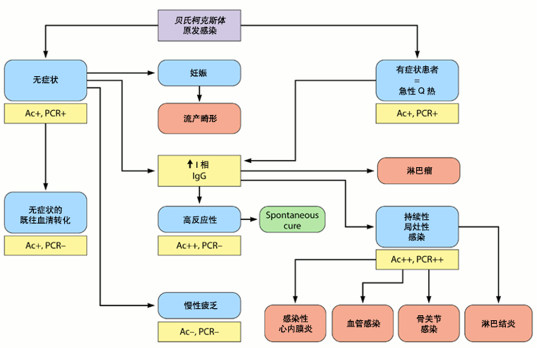 [Figure caption and citation for the preceding image starts]: 贝氏柯克斯体自然病程和血清学/PCR 结果。Ac+:贝氏柯克斯体抗体阳性;Ac++:贝氏柯克斯体抗体强阳性;Ac-:贝氏柯克斯体抗体阴性;PCR+:贝氏柯克斯体 PCR 阳性;PCR++:贝氏柯克斯体 PCR 强阳性; PCR-:贝氏柯克斯体 PCR 阴性Eldin C, et al. Clin Microbiol Rev 2016;经许可后使用 [Citation ends].
因为血清转化可能延迟长达 6 周,如果临床表现符合,但初次血清学检查为阴性,必须在第 15、30 和 45 天进行血清学对照检查。通过冻干法可以增加血液/血清 PCR 的敏感性,[68]Edouard S, Raoult D. Lyophilization to improve the sensitivity of qPCR for bacterial DNA detection in serum: the Q fever paradigm. J Med Microbiol. 2016;65:462-467.http://www.ncbi.nlm.nih.gov/pubmed/27008653?tool=bestpractice.com在上述情况下可能非常有用。
组织活检、免疫组化、 [Figure caption and citation for the preceding image starts]: 贝氏柯克斯体骨炎:免疫组化:棕色显示出单核细胞/巨噬细胞中的细菌Hubert Lepidi, Institut Hospitalo-Universitaire Méditerranée Infection [Citation ends].
 [Figure caption and citation for the preceding image starts]: 贝氏柯克斯体肺纤维化:免疫组化;棕色染色显示出单核细胞/巨噬细胞中的细菌Hubert Lepidi, Institut Hospitalo-Universitaire Méditerranée Infection [Citation ends].
 [Figure caption and citation for the preceding image starts]: 贝氏柯克斯体心内膜炎:免疫组化。图片提示,炎症水平低。棕色染色显示出单核细胞/巨噬细胞内的细菌。通常没有赘生物Hubert Lepidi, Institut Hospitalo-Universitaire Méditerranée Infection [Citation ends].
 [Figure caption and citation for the preceding image starts]: 贝氏柯克斯体慢性淋巴结炎:免疫组化。图像显示,淋巴结中孤立的受感染细胞(单核细胞/巨噬细胞)。棕色染色显示出单核细胞/巨噬细胞中的细菌Hubert Lepidi, Institut Hospitalo-Universitaire Méditerranée Infection [Citation ends].
 [Figure caption and citation for the preceding image starts]: 心内膜炎患者的贝氏柯克斯体慢性肝炎:免疫组化。图像显示,不存在急性 Q 热中常见的甜甜圈状肉芽肿。棕色染色显示出单核细胞/巨噬细胞中的细菌Hubert Lepidi, Institut Hospitalo-Universitaire Méditerranée Infection [Citation ends].和荧光原位杂交技术 (FISH)
 [Figure caption and citation for the preceding image starts]: 贝氏柯克斯体肺假性肿瘤:荧光原位杂交技术 (FISH)。红色:免疫荧光 (IF)。绿色:使用特异性 16S rRNA 探针的 FISH。黄色:IF 和 FISH 共定位确认肺假性肿瘤的 2 个细胞细胞质中存在细菌Gilles Audoly, Institut Hospitalo-Universitaire Méditerranée Infection [Citation ends].是金标准,但仅在专业实验室进行这些检查。免疫组化的优势是能够识别细胞类型,而 FISH 的敏感性远高于免疫组化。由于有淋巴瘤风险,在慢性淋巴结炎(根据 18F 氟脱氧葡萄糖 [FDG] PET/CT)患者中,应当常规进行淋巴结活检。[11]Melenotte C, Million M, Audoly G, et al. B-cell non-Hodgkin lymphoma linked to Coxiella burnetii. Blood. 2016;127:113-121.http://www.ncbi.nlm.nih.gov/pubmed/26463422?tool=bestpractice.com
影像学
CXR:
如怀疑肺部并发症,在急性疾病中可能需要此项检查。 [Figure caption and citation for the preceding image starts]: 贝氏柯克斯体肺炎。一名 21 岁贝氏柯克斯体肺炎女性的胸部 X 射线和 CT 扫描;CXR 显示双侧中下肺野多个区域存在软组织实变;CT 扫描显示边界不清的小叶中央型结节和气腔实变Okimoto N, et al. Respirology.2004;9:278-282;经 John Wiley & Sons 有限公司许可后使用 [Citation ends].结果可能从正常到双肺多发性非系统化(无明显的规律或分布特征)阴影,最符合非典型肺炎特征。[5]Maurin M, Raoult D. Q fever. Clin Microbiol Rev. 1999;12:518-553.http://cmr.asm.org/content/12/4/518.fullhttp://www.ncbi.nlm.nih.gov/pubmed/10515901?tool=bestpractice.com胸部 X 线表现最常见的异常是节段性或大叶性阴影。[Figure caption and citation for the preceding image starts]: 贝氏柯克斯体肺炎。一名 21 岁贝氏柯克斯体肺炎女性的胸部 X 射线和 CT 扫描;CXR 显示双侧中下肺野多个区域存在软组织实变;CT 扫描显示边界不清的小叶中央型结节和气腔实变Okimoto N, et al. Respirology.2004;9:278-282;经 John Wiley & Sons 有限公司许可后使用 [Citation ends].多个圆形阴影是 Q 热肺炎的特征,但不常见。胸腔积液罕见。在持续性局灶性感染患者中,CXR 可能发现两种不同的持续性局灶性感染:间质性纤维化和肺假性肿瘤。
超声心动图:
在急性感染期间常规建议经胸超声心动图 (TTE) 检查,用于排除无症状但可能需要抗生素预防治疗的潜在心脏病变。[3]Eldin C, Mélenotte C, Mediannikov O, et al. From Q fever to Coxiella burnetii infection: a paradigm change. Clin Microbiol Rev. 2017;30:115-190.http://www.ncbi.nlm.nih.gov/pubmed/27856520?tool=bestpractice.com在 IgG 抗心磷脂抗体水平非常高的急性心内膜炎患者中,可经常发现大的短暂赘生物。[9]Million M, Thuny F, Bardin N, et al. Antiphospholipid antibody syndrome with valvular vegetations in acute Q fever. Clin Infect Dis. 2016;62:537-544.http://www.ncbi.nlm.nih.gov/pubmed/26585519?tool=bestpractice.com在慢性心内膜炎患者中,赘生物小或不存在。[69]Million M, Raoult D. The pathogenesis of the antiphospholipid syndrome. N Engl J Med. 2013;368:2335.http://www.ncbi.nlm.nih.gov/pubmed/23758255?tool=bestpractice.com经常有结节性病变和钙化。
在年龄>40 岁、有急性感染但 TTE 为阴性、IgG aCL 抗体≥75 GPLU 的患者中,建议进行经食管超声心动图 (TEE) 检查,用于识别心脏病变。
肝脏超声:
胸部和脑部 CT 扫描:
腹部 CT 扫描或超声:
在年龄>65 岁并且目前吸烟或既往吸烟或有动脉瘤家族史的急性感染患者中,建议进行这些检查。主动脉瘤患者如有急性感染,需要特异性治疗方法。[3]Eldin C, Mélenotte C, Mediannikov O, et al. From Q fever to Coxiella burnetii infection: a paradigm change. Clin Microbiol Rev. 2017;30:115-190.http://www.ncbi.nlm.nih.gov/pubmed/27856520?tool=bestpractice.com
18F-FDG PET/CT 影像:
对识别持续性局灶性感染很关键。[70]Eldin C, Melenotte C, Million M, et al. 18F-FDG PET/CT as a central tool in the shift from chronic Q fever to Coxiella burnetii persistent focalized infection: a consecutive case series. Medicine (Baltimore). 2016;95:e4287.http://www.ncbi.nlm.nih.gov/pubmed/27559944?tool=bestpractice.com
它能够显示心内膜炎、血管感染、淋巴结炎和骨关节感染,不使用这种技术,无法发现这些病变。 [Figure caption and citation for the preceding image starts]: 通过 PET 扫描诊断的 Q 热心内膜炎:18F-氟脱氧葡萄糖 PET/CT。在这名有心脏瓣膜疾病病史并且血清学结果升高的无症状患者中,PET 扫描诊断为自体瓣膜主动脉心内膜炎伴有胸主动脉和腰主动脉霉菌性动脉瘤Institut Hospitalo-Universitaire Méditerranée Infection(患者同意使用) [Citation ends].
 [Figure caption and citation for the preceding image starts]: 通过 PET 扫描诊断的 Q 热霉菌性胸主动脉瘤:18F-氟脱氧葡萄糖 PET/CT。在这名有心脏瓣膜疾病病史并且血清学结果升高的无症状患者中,PET 扫描诊断为自体瓣膜主动脉心内膜炎伴有胸主动脉和腰主动脉霉菌性动脉瘤Institut Hospitalo-Universitaire Méditerranée Infection(患者同意使用) [Citation ends].
 [Figure caption and citation for the preceding image starts]: 通过 PET 扫描诊断的 Q 热霉菌性腰主动脉瘤:18F-氟脱氧葡萄糖 PET/CT。在这名有心脏瓣膜疾病病史并且血清学结果升高的无症状患者中,PET 扫描诊断为自体瓣膜主动脉心内膜炎伴有胸主动脉和腰主动脉霉菌性动脉瘤Institut Hospitalo-Universitaire Méditerranée Infection(患者同意使用) [Citation ends].
如果患者有持续性症状和/或血清学检查持续升高和/或血液/血清或任何样本的 PCR 阳性且临床表现不符合原发感染,则此项检查是标准解剖结构检查的一部分。对于具有下列情况的急性感染患者,尤其推荐此项检查:[3]Eldin C, Mélenotte C, Mediannikov O, et al. From Q fever to Coxiella burnetii infection: a paradigm change. Clin Microbiol Rev. 2017;30:115-190.http://www.ncbi.nlm.nih.gov/pubmed/27856520?tool=bestpractice.com
持续存在 I 相 IgG≥1:800 和/或临床进展不良的征象[Figure caption and citation for the preceding image starts]: 通过 PET 扫描诊断的 Q 热心内膜炎:18F-氟脱氧葡萄糖 PET/CT。在这名有心脏瓣膜疾病病史并且血清学结果升高的无症状患者中,PET 扫描诊断为自体瓣膜主动脉心内膜炎伴有胸主动脉和腰主动脉霉菌性动脉瘤Institut Hospitalo-Universitaire Méditerranée Infection(患者同意使用) [Citation ends].[Figure caption and citation for the preceding image starts]: 通过 PET 扫描诊断的 Q 热霉菌性胸主动脉瘤:18F-氟脱氧葡萄糖 PET/CT。在这名有心脏瓣膜疾病病史并且血清学结果升高的无症状患者中,PET 扫描诊断为自体瓣膜主动脉心内膜炎伴有胸主动脉和腰主动脉霉菌性动脉瘤Institut Hospitalo-Universitaire Méditerranée Infection(患者同意使用) [Citation ends].[Figure caption and citation for the preceding image starts]: 通过 PET 扫描诊断的 Q 热霉菌性腰主动脉瘤:18F-氟脱氧葡萄糖 PET/CT。在这名有心脏瓣膜疾病病史并且血清学结果升高的无症状患者中,PET 扫描诊断为自体瓣膜主动脉心内膜炎伴有胸主动脉和腰主动脉霉菌性动脉瘤Institut Hospitalo-Universitaire Méditerranée Infection(患者同意使用) [Citation ends].
血管移植或动脉瘤病史
原因不明的血清学检查结果(I 相 IgG≥1:800)或临床怀疑持续性感染。
它还可用于识别有血管假体和/或动脉瘤患者中的感染,以及识别需要手术切除受感染血管组织的患者。
下面的流程为管理急性 Q 热提供了一个循序渐进的方法:[3]Eldin C, Mélenotte C, Mediannikov O, et al. From Q fever to Coxiella burnetii infection: a paradigm change. Clin Microbiol Rev. 2017;30:115-190.http://www.ncbi.nlm.nih.gov/pubmed/27856520?tool=bestpractice.com
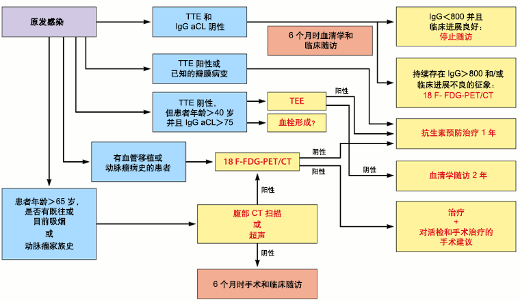 [Figure caption and citation for the preceding image starts]: TTE:经胸超声心动图;IgG aCL:IgG 抗心磷脂抗体;18 F FDG-PET/CT:18F-氟脱氧葡萄糖正电子发射断层成像联合计算机断层扫描Eldin C, et al. Clin Microbiol Rev 2016;经许可后使用 [Citation ends].