胸腰段椎体骨折的诊断主要是基于损伤机制和临床症状。 不管是采用非手术治疗还是手术治疗,都需要采用适当的影像学技术进一步评估和制定治疗计划。 应注意避免对多发性损伤患者的胸腰段椎体骨折的漏诊。 胸腰段椎体骨折的漏诊率高达19%。[27]Dai LY, Yao WF, Cui YM, et al. Thoracolumbar fractures in patients with multiple injuries: diagnosis and treatment-a review of 147 cases. J Trauma. 2004 Feb;56(2):348-55.http://www.ncbi.nlm.nih.gov/pubmed/14960979?tool=bestpractice.com
对于意识受损的患者,需要患者的亲友、护理人员、护士或医生,或者其他与患者接触过的人提供观察资料。
获取高质量的病史对找出患者的受伤机制(如高空跌落、交通事故、运动受伤)具有重要意义,可能提示受伤的严重程度。 检查是否存在脊柱梯状畸形和疼痛。 脊柱无疼痛或变形,包括神经功能障碍,则表明可能无胸腰段椎体骨折。 但是,如果脊椎疼痛或变形,则只能进一步通过成像技术(MRI、CT)确认脊椎是否骨折或受到其他损伤(如韧带损伤、血肿、椎旁软组织损伤)。
初步评估和复苏
现场初步评估包括:[28]American College of Surgeons. Advanced trauma life support manual, 7th ed. Chicago, IL: American College of Surgeons; 2004.[29]National Institute for Health and Care Excellence. Spinal injury: assessment and initial management. February 2016. https://www.nice.org.uk [internet publication].https://www.nice.org.uk/guidance/ng41
通过颈椎固定维持气道通畅
呼吸和通气(评估呼吸率、呼吸深度以及氧饱和度;呼吸浅促可能表明更高位置的颈椎骨折)
采用出血控制的循环(评估心率、血压和毛细血管再充盈时间;如果持续出血,则使用低血压复苏策略来将目标平均动脉血压维持在 50 到 65 mmHg)。[30]Bickell WH, Wall MJ Jr, Pepe PE, et al. Immediate versus delayed fluid resuscitation for hypotensive patients with penetrating torso injuries. N Engl J Med. 1994 Oct 27;331(17):1105-9.http://www.nejm.org/doi/full/10.1056/NEJM199410273311701#t=articlehttp://www.ncbi.nlm.nih.gov/pubmed/7935634?tool=bestpractice.com[31]Morrison CA, Carrick MM, Norman MA, et al. Hypotensive resuscitation strategy reduces transfusion requirements and severe postoperative coagulopathy in trauma patients with hemorrhagic shock: preliminary results of a randomized controlled trial. J Trauma. 2011 Mar;70(3):652-63.http://www.ncbi.nlm.nih.gov/pubmed/21610356?tool=bestpractice.com 为改善灌注,在急性脊髓损伤后最初 7 天,平均动脉压应维持在 85 至 90 mmHg[32]Blood pressure management after acute spinal cord injury. Neurosurgery. 2002 Mar;50(3 Suppl):S58-62.http://www.ncbi.nlm.nih.gov/pubmed/12431288?tool=bestpractice.com
残疾(评估格拉斯哥昏迷评分、双边瞳孔大小和反应以及单侧体征)
对患者进行露体检查,查看是否有明显的重伤部位。
如果怀疑有急性脊髓损伤(伴有或不伴有脊柱损伤),应当先将患者转移至最近的创伤中心,确定和治疗威胁生命的疾病,然后再转移至脊髓损伤中心。[29]National Institute for Health and Care Excellence. Spinal injury: assessment and initial management. February 2016. https://www.nice.org.uk [internet publication].https://www.nice.org.uk/guidance/ng41 在脊髓损伤后的急性期内,静脉使用吗啡为缓解疼痛的一线药物。 静脉使用氯胺酮为二线药物;如果获得静脉使用通路有延迟,则也可以使用鼻内给药氯胺酮或二乙酰吗啡(如有)。
病史
需要重点记录脊柱疼痛、瘫痪或感觉异常的详细病史。 例如,在瘫痪或完全麻木以前患者报告了身体受累部位的任何运动和感觉,则应查明该事件发生的时间,这一点非常重要。 对于疼痛,患者所述的背痛程度通常与其脊柱损伤程度有关。
在必须进行直线式脊柱固定的胸椎或腰骶椎损伤评估期间,重大发现包括:[29]National Institute for Health and Care Excellence. Spinal injury: assessment and initial management. February 2016. https://www.nice.org.uk [internet publication].https://www.nice.org.uk/guidance/ng41
年龄 ≥ 65 岁并有胸椎或腰骶椎疼痛
严重损伤的警示性特征(例如从>3 m [>10 英尺] 的地方跌落;[33]Velmahos GC, Spaniolas K, Alam HB, et al. Falls from height: spine, spine, spine! J Am Coll Surg. 2006 Nov;203(5):605-11.http://www.ncbi.nlm.nih.gov/pubmed/17084320?tool=bestpractice.com 摔下时脚或者臀部先着地;骑马事故;速度>100 km/h [>60 英里/小时] 时发生机动车事故 [motor vehicle accident, MVA];从机动车中弹出的事故;患者躯干前面有安全带印迹的机动车事故 [与腰椎 Chance 骨折相关];背部疼痛、下肢神经功能障碍 [虚弱或麻痹、麻木] 或括约肌功能障碍)
异常神经系统体征(运动受限/感觉缺乏);(触诊时)脊柱产生新的畸形或者骨中线压痛;咳嗽时中线或者脊柱疼痛
坐下、站立或踏步时疼痛或者异常神经系统体征(如出现疼痛停止动作)
脊柱的另一个区域疑似脊柱骨折
既往存在脊柱病理学改变。
如果轻微创伤引起低能损伤,则需排除骨质疏松症或其他恶性原因(例如肿瘤和代谢疾病)。另外,既往病史可能提示一些危险因素,包括合并骨质疏松症、既往椎体骨折、潜在肿瘤(例如多发性骨髓瘤、肿瘤骨转移)或潜在代谢性或炎症性疾病(例如成骨不全症、骨质疏松症、类风湿性关节炎和强直性脊柱炎)。 [Figure caption and citation for the preceding image starts]: 腰椎磁共振成像:矢状位(T2加权序列)显示T12椎体有骨质疏松性骨折来自B. Nurboja博士和D. Choi先生个人收集的资料 [Citation ends].
 [Figure caption and citation for the preceding image starts]: 胸椎磁共振成像:矢状位(T2加权序列)显示T10椎体有多发性骨髓瘤造成的病理性骨折来自B. Nurboja博士和D. Choi先生个人收集的资料 [Citation ends].
体格检查
对于失去意识的患者,应评估能够表明其脊髓功能水平的征象:
对于有意识的患者,需要进行视诊、触诊和全面的神经系统检查。
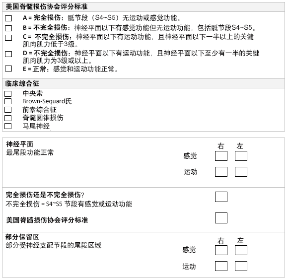 [Figure caption and citation for the preceding image starts]: 美国脊柱损伤协会脊髓损伤神经学分型标准美国脊柱损伤协会,经许可后使用 [Citation ends].
应注意是否存在瘀血。 脊椎的畸形情况(梯状变形或肿胀)能够表明损伤的严重程度。 对脊椎进行触诊检查时,引起的触痛程度与脊柱损伤的程度相关。
急性麻木或感觉异常可能是由脊髓或神经根受到压迫引起的。 对神经系统损伤水平,包括感觉的皮节分布进行仔细的临床评估必不可少,所以就要更好地分析脊椎X线片。 神经损伤水平是指保留身体两侧正常感觉功能的最尾端的脊髓节段水平。 对单个皮节的左右两侧感觉功能同时进行评估有助于提高感觉功能诊断的敏感性。
肌肉痉挛表明上运动神经元病变,张力减低或迟缓表明脊髓休克(暂时性休克)或下运动神经元病变。 踝阵挛是指用力使踝关节背屈后,踝关节不由自主地节律性伸屈运动3次以上,即为阵挛异常。 阵挛异常表明上运动神经元病变。
上运动神经元病变的其他体征包括与颈椎脊髓压迫相关的霍夫曼征(轻弹中指甲床造成其他手指突然向同侧抽搐)和巴宾斯基征阳性(击打脚的侧面或跖面时,拇趾伸张,其余脚趾向同侧外展)。
肛门括约肌反射损失表明脊髓受伤,但这并非某一特定脊髓水平损伤才出现的特定现象。
如果存在神经功能障碍,则需要诱发球海绵体肌反射(S3-S4)。 对于男性,要挤压龟头;对于女性,要按压阴蒂,并感觉到肛门括约肌收缩。 无此反射表明脊髓休克、骶段脊髓损伤或骶神经根损伤。
尿失禁通常是由高位损伤造成脊髓损害引起的。 下行纤维的抑制性调控受损造成膀胱容量缩小,进而导致尿失禁(高张性膀胱)。 另外,高位损伤可能在急性期导致尿潴留和充溢性尿失禁,而且在完全性损伤时无痛感(即患者感觉不到膀胱充盈)。 持续的,无痛性尿储留表明影响膀胱神经支配的马尾受伤,造成尿储留。
15%至20%的脊髓损伤患者为多节段脊柱骨折。[11]Vaccaro AR, An HS, Lin S, et al. Noncontiguous injuries of the spine. J Spinal Disord. 1992 Sep;5(3):320-9.http://www.ncbi.nlm.nih.gov/pubmed/1520991?tool=bestpractice.com 脊髓损伤患者通常还有其他相关损伤,例如合并多系统损伤 (80%)[11]Vaccaro AR, An HS, Lin S, et al. Noncontiguous injuries of the spine. J Spinal Disord. 1992 Sep;5(3):320-9.http://www.ncbi.nlm.nih.gov/pubmed/1520991?tool=bestpractice.com 和头部损伤 (41%)。[15]Davidoff G, Roth E, Morris J, et al. Assessment of closed head injury in trauma related spinal cord injury. Paraplegia. 1986 Apr;24(2):97-104.http://www.ncbi.nlm.nih.gov/pubmed/3714296?tool=bestpractice.com
脊髓休克与神经性休克
脊髓受伤后,脊髓的神经纤维可能会立即除极,然后再复极。[23]Kakulas BA. Pathology of spinal injuries. Cent Nerv Syst Trauma. 1984 Winter;1(2):117-29.http://www.ncbi.nlm.nih.gov/pubmed/6545680?tool=bestpractice.com
根据受伤后72小时或1周后的神经系统检查来预测功能康复会更可靠。[34]Brown PJ, Marino RJ, Herbison GJ, et al. The 72-hour examination as a predictor of recovery in motor complete quadriplegia. Arch Phys Med Rehabil. 1991 Jul;72(8):546-8.http://www.ncbi.nlm.nih.gov/pubmed/2059130?tool=bestpractice.com
通常情况下,在决定是否采取手术治疗时,并不认为脊髓休克是一种限制因素。如果患者需要接受紧急脊髓减压,等待 72 小时至脊髓休克完全消失后再进行手术可能产生不利影响。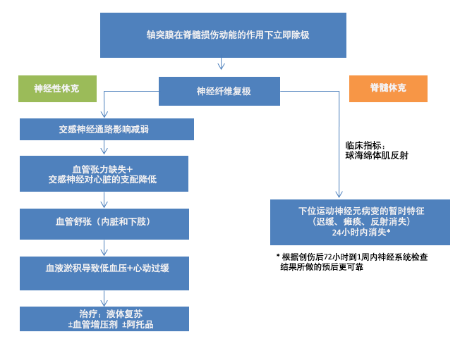 [Figure caption and citation for the preceding image starts]: 脊髓损伤B. Nurboja博士创建 [Citation ends].
成年人影像学检查
对于清醒且反应灵敏的患者,如果没有临床证据表明有胸腰段椎体损伤,则不需要拍摄常规胸腰椎 X 线。[35]Durham RM, Luchtefeld WB, Wibbenmeyer L, et al. Evaluation of the thoracic and lumbar spine after blunt trauma. Am J Surg. 1995 Dec;170(6):681-4.http://www.ncbi.nlm.nih.gov/pubmed/7492026?tool=bestpractice.com 急性脊柱损伤中如果出现以下情况,应拍摄胸腰椎片:1)背痛或中线压痛;2)胸腰损伤局部体征(如淤青);3)异常神经系统体征;4)颈椎骨折,5)格拉斯哥昏迷评分<15;6)重大牵张性损伤;7)酒精或药物中毒。[36]Hsu JM, Joseph T, Ellis AM. Thoracolumbar fracture in blunt trauma patients: guidelines for diagnosis and imaging. Injury. 2003 Jun;34(6):426-33.http://www.ncbi.nlm.nih.gov/pubmed/12767788?tool=bestpractice.com 脊椎的一个节段发生骨折,提示其他节段发生脊柱骨折的风险增高。 因此,要确诊某一节段脊椎是否发生骨折,可能表明有必要对其他脊椎节段也进行检查。[37]American College of Radiology. ACR appropriateness criteria: suspected spine trauma. 2012 [internet publication].https://acsearch.acr.org/docs/69359/Narrative/
外伤时拍摄胸部和腹部 X 线并不能够诊断椎骨骨折和椎体排列情况。[38]Bolesta MJ, Bohlman HH. Mediastinal widening associated with fractures of the upper thoracic spine. J Bone Joint Surg Am. 1991 Mar;73(3):447-50.http://www.ncbi.nlm.nih.gov/pubmed/2002082?tool=bestpractice.com 如果怀疑胸腰段椎体骨折,至少需要拍摄脊椎的前后位以及侧位片。[39]Keene JS. Radiographic evaluation of thoracolumbar fractures. Clin Orthop Relat Res. 1984 Oct;(189):58-64.http://www.ncbi.nlm.nih.gov/pubmed/6478704?tool=bestpractice.com[40]Ballock RT, Mackersie R, Abitbol JJ, et al. Can burst fractures be predicted from plain radiographs? J Bone Joint Surg Br. 1992 Jan;74(1):147-50.http://www.bjj.boneandjoint.org.uk/content/jbjsbr/74-B/1/147.full.pdfhttp://www.ncbi.nlm.nih.gov/pubmed/1732246?tool=bestpractice.com[41]Valentini MC, Busch R, Ferraris MM, et al. The role of imaging in the choice of correct treatment of unstable thoraco-lumbar fractures. Eur J Radiol. 2006 Sep;59(3):331-5.http://www.ncbi.nlm.nih.gov/pubmed/16930904?tool=bestpractice.com
前后位片可以看到椎体高度变矮、横向直径和基底间距增大、棘突间的空隙变宽、关节突关节分离和侧向平移。
侧位片可以看到上终板骨折、前楔形变形、前壁皮层不规则以及椎体错位。
在30%的病例中,普通X光片无法显示椎体后皮质受累的情况,从而导致将爆裂性骨折误诊为楔形骨折。 为此,普通平片上显示楔形骨折或可疑压缩性骨折时,建议进行CT扫描以确诊。
如果怀疑患儿受到身体虐待,临床医生应考虑胸腰段椎体骨折的可能性。 一项针对受虐待的儿童进行的研究发现,1%的非意外头部损伤与脊椎损伤相关,1%至3%的受虐儿童骨折属于椎骨骨折。[42]Merten DF, Radkowski MA, Leonidas JC. The abused child: a radiological reappraisal. Radiology. 1983 Feb;146(2):377-81.http://www.ncbi.nlm.nih.gov/pubmed/6849085?tool=bestpractice.com 在对19篇儿童脊椎损伤文献的系统回顾中发现,其纳入的25名患儿中有23人不到2岁,12人有胸腰段椎体骨折,其中75%为骨折脱位,25%为脊椎压缩性骨折。 系统评价建议所有2岁以下疑似非意外损伤的患儿都应拍摄胸腰椎 X 线检查。[43]Kemp AM, Joshi AH, Mann M, et al. What are the clinical and radiological characteristics of spinal injuries from physical abuse: a systematic review. Arch Dis Child. 2010 May;95(5):355-60.http://www.ncbi.nlm.nih.gov/pubmed/19946011?tool=bestpractice.com
由于CT扫描能够更好地显现椎弓、关节突关节和椎管,在病情稳定的前提下,所有可疑的胸腰段椎体损伤患者都应进行脊椎CT扫描。[44]Keene JS, Goletz TH, Lilleas F, et al. Diagnosis of vertebral fractures: a comparison of conventional radiography, conventional tomography, and computed axial tomography. J Bone Joint Surg Am. 1982 Apr;64(4):586-94.http://www.ncbi.nlm.nih.gov/pubmed/7068700?tool=bestpractice.com 但是,如果已经拍摄了普通X线片,则在出现以下症状时,应拍摄CT片进行脊椎评估:
椎弓根间距增大(爆裂性骨折特征)[45]Martijn A, Veldhuis EF. The diagnostic value of interpediculate distance assessment on plain films in thoracic and lumbar spine injuries. J Trauma. 1991 Oct;31(10):1393-5.http://www.ncbi.nlm.nih.gov/pubmed/1942150?tool=bestpractice.com
横型骨折(可能与其他脊椎或骨盆重大损伤相关)。[46]Krueger MA, Green DA, Hoyt D, et al. Overlooked spine injuries associated with lumbar transverse process fractures. Clin Orthop Relat Res. 1996 Jun;(327):191-5.http://www.ncbi.nlm.nih.gov/pubmed/8641063?tool=bestpractice.com
由于一些疾病(如硬膜外血肿或椎间盘突出造成脊髓受压)只在磁共振成像上显示出来,即使脊椎的CT片或X线片显示正常也可能存在神经功能障碍,因此需要对脊椎进行磁共振成像扫描。
脊柱 MRI 可确定髓内病变(如外伤后囊肿、血肿和水肿)和髓外压迫(如椎间盘、血肿和骨折碎片),并且可用于评估后方韧带复合体。[7]Ferguson RL, Allen BL Jr. A mechanistic classification of thoracolumbar spine fractures. Clin Orthop Related Res. 1984 Oct;(189):77-88.http://www.ncbi.nlm.nih.gov/pubmed/6478706?tool=bestpractice.com
如果未进行MRI脊椎扫描或患者不适合接受该项检查(例如患者装有金属植入物,血流动力学不稳定或严重的幽闭恐怖症),则可能需要进行CT脊髓造影术。
全身 CT(包括从头顶至脚趾的扫描图、然后进行从头顶至大腿中部的 CT )适用于有多处损伤及疑似脊柱损伤的成年人。[29]National Institute for Health and Care Excellence. Spinal injury: assessment and initial management. February 2016. https://www.nice.org.uk [internet publication].https://www.nice.org.uk/guidance/ng41[Figure caption and citation for the preceding image starts]: 胸椎磁共振成像:矢状位(T2加权序列)显示T10椎体有多发性骨髓瘤造成的病理性骨折来自B. Nurboja博士和D. Choi先生个人收集的资料 [Citation ends]. [Figure caption and citation for the preceding image starts]: 腰椎磁共振成像:轴位(T2加权序列)显示T12椎体有骨质疏松性骨折来自B. Nurboja博士和D. Choi先生个人收集的资料 [Citation ends].[Figure caption and citation for the preceding image starts]: 腰椎磁共振成像:矢状位(T2加权序列)显示T12椎体有骨质疏松性骨折来自B. Nurboja博士和D. Choi先生个人收集的资料 [Citation ends].
儿童影像学检查(年龄<16 岁)
如果初始 X 线检查异常,则有疑似胸腰段椎体损伤的儿童应当进行 CT 检查。 对于疑似有颈椎损伤的儿童首选 MRI 而非 CT。 如果确定脊柱存在新骨折,则应该也对脊柱其他部分进行影像学检查。[29]National Institute for Health and Care Excellence. Spinal injury: assessment and initial management. February 2016. https://www.nice.org.uk [internet publication].https://www.nice.org.uk/guidance/ng41
如果需要进一步放射评估,则应该使用临床判断来减少对身体区域进行 CT 检查,因为儿童暴露于 CT 扫描后患有的癌症风险(包括甲状腺癌)会增加。[29]National Institute for Health and Care Excellence. Spinal injury: assessment and initial management. February 2016. https://www.nice.org.uk [internet publication].https://www.nice.org.uk/guidance/ng41[47]Mathews JD, Forsythe AV, Brady Z, et al. Cancer risk in 680,000 people exposed to computed tomography scans in childhood or adolescence: data linkage study of 11 million Australians. BMJ. 2013 May 21;346:f2360http://www.ncbi.nlm.nih.gov/pmc/articles/PMC3660619/http://www.ncbi.nlm.nih.gov/pubmed/23694687?tool=bestpractice.com
骨折分型
建议按照公认的分型体系划分骨折,如Denis或AO分型系统。[1]Gertzbein SD. Spine update: classification of thoracic and lumbar fractures. Spine (Phila Pa 1976). 1994 Mar 1;19(5):626-8.http://www.ncbi.nlm.nih.gov/pubmed/8184361?tool=bestpractice.com[2]Denis F. The three-column spine and its significance in the classification of acute thoracolumbar spinal injuries. Spine (Phila Pa 1976). 1983 Nov-Dec;8(8):817-31.http://www.ncbi.nlm.nih.gov/pubmed/6670016?tool=bestpractice.com 这两种分型系统均是非常有效的分型体系,对于非手术或手术治疗规划有帮助。 AO分型更为常用,因为它提供了一种全面的分型方法,描述了损伤的性质、不稳定程度和预后等,这些对选择最适当的疗法具有重要意义。  [Figure caption and citation for the preceding image starts]: 改良Maegerl(AO/ASIF)胸腰段椎体骨折分型作者摘自:Gertzbein SD. Spine update: classification of thoracic and lumbar fractures. Spine. 1994;19:626-628 [Citation ends].
骨折稳定性评估
进一步的治疗方案取决于骨折稳定性的评估结果。
脊椎不稳定的定义为“在生理负荷下,脊椎应能维持各椎骨的相关关系,使脊髓或其神经根不受损害或刺激,也不会发生变形或疼痛。如果脊椎丧失这种能力,则称为脊椎不稳定。”[48]Panjabi MM, Thibodeau LL, Crisco JJ 3rd, et al. What constitutes spinal instability? Clin Neurosurg. 1988;34:313-39.http://www.ncbi.nlm.nih.gov/pubmed/3288400?tool=bestpractice.com
讨论骨折不稳定性时,需要考虑2个主要概念:
骨折稳定性能够通过拍摄胸腰区域的MRI片确定,MRI片能显示韧带复合体断裂的情况。