临床上治疗持续性肺部浸润时需要评估宿主因素(年龄、合并症、免疫缺陷)、症状严重程度和非感染性病因的可能性。采集病史并进行临床检查,同时辅以实验室评估和放射影像学技术,可以帮助缩小鉴别诊断的范围。[15]Woodhead M, Blasi F, Ewig S, et al; Joint Taskforce of the European Respiratory Society and European Society for Clinical Microbiology and Infectious Diseases. Guidelines for the management of adult lower respiratory tract infections. Clin Microbiol Infect. 2011 Nov;17 (Suppl 6):E1-59.https://www.escmid.org/fileadmin/src/media/PDFs/4ESCMID_Library/2Medical_Guidelines/ESCMID_Guidelines/Woodhead_et_al_CMI_Sep_2011_LRTI_GL_fulltext.pdfhttp://www.ncbi.nlm.nih.gov/pubmed/21951385?tool=bestpractice.com[21]Low DE, Mazzulli T, Marrie T. Progressive and nonresolving pneumonia. Curr Opin Pulm Med. 2005 May;11(3):247-52.http://www.ncbi.nlm.nih.gov/pubmed/15818188?tool=bestpractice.com[22]Weyers CM, Leeper KV. Nonresolving pneumonia. Clin Chest Med. 2005 Mar;26(1):143-58.http://www.ncbi.nlm.nih.gov/pubmed/15802176?tool=bestpractice.com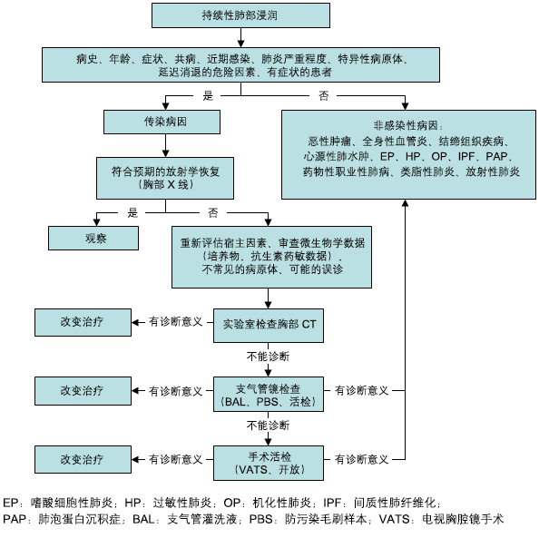 [Figure caption and citation for the preceding image starts]: 持续性肺部浸润影诊断路径的图表由 Athanasia Pataka 创建 [Citation ends].
病史
发热和咳痰提示感染性疾病的可能性,然而存在持续性肺部浸润影的患者可能仅有影像学表现而无临床症状。肺部感染(尤其导致影像学上延迟缓解的病原体感染)病史可能预示存在延缓吸收性肺炎。
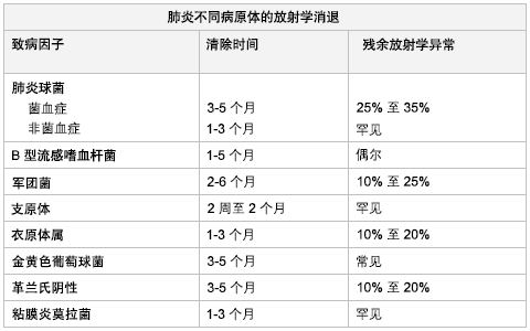 [Figure caption and citation for the preceding image starts]: 致病病原体与肺炎的影像学吸收由 Athanasia Pataka 创建 [Citation ends]. 合并症(COPD、酗酒、肾衰竭)可能也会延迟肺炎的影像学缓解。[5]Mittl RL Jr, Schwab RJ, Duchin JS, et al. Radiographic resolution of community-acquired pneumonia. Am J Respir Crit Care Med. 1994 Mar;149(3 Pt 1):630-5.http://www.ncbi.nlm.nih.gov/pubmed/8118630?tool=bestpractice.com[7]El Solh AA, Aquilina AT, Gunen H, et al. Radiographic resolution of community-acquired bacterial pneumonia in the elderly. J Am Geriatr Soc. 2004 Feb;52(2):224-9.http://www.ncbi.nlm.nih.gov/pubmed/14728631?tool=bestpractice.com 免疫抑制患者最初必须进行感染的评估。在无感染性病因的情况下,则必须考虑非感染性病因,如放射、药物反应和弥漫性肺泡出血。
20%的持续性肺部浸润影是由非感染性病因引起。[13]Menendez R, Torres A, Zalacaín R, et al. Neumofail Group. Risk factors of treatment failure in community acquired pneumonia: implications for disease outcome. Thorax. 2004 Nov;59(11):960-5.http://www.pubmedcentral.nih.gov/picrender.fcgi?artid=1746855&blobtype=pdfhttp://www.ncbi.nlm.nih.gov/pubmed/15516472?tool=bestpractice.com 某些持续性浸润影可能与心肌梗死、心绞痛和外周水肿的病史相关。
血尿可能提示肺泡出血综合征,而关节疼痛或皮疹可能提示结缔组织病。
哮喘和持续的游走性浸润影可能提示过敏性支气管肺曲霉病。
用药史十分重要,因为药物(例如胺碘酮、博来霉素、环磷酰胺、长春新碱、紫杉醇)可能会导致肺浸润。
体格检查
表现为持续性肺部浸润影的患者可能不一定有典型的肺部体征(如听诊捻发音和啰音、胸部叩诊浊音)。 然而,对于缓慢吸收性或不吸收性肺炎患者应该与其他重症患者一样进行全面的检查。
对于免疫功能正常的肺炎患者,对抗生素治疗的临床反应是持续性肺部浸润是否进行下一步诊断性检查最为重要的因素。[4]Mandell LA, Wunderink RG, Anzueto A, et al. Infectious Diseases Society of America/American Thoracic Society consensus guidelines on the management of community-acquired pneumonia in adults. Clin Infect Dis. 2007 Mar 1;44 (Suppl 2):S27-72.https://academic.oup.com/cid/article/44/Supplement_2/S27/372079http://www.ncbi.nlm.nih.gov/pubmed/17278083?tool=bestpractice.com[14]Menendez R, Torres A. Treatment failure in community-acquired pneumonia. Chest. 2007 Oct;132(4):1348-55.http://www.ncbi.nlm.nih.gov/pubmed/17934120?tool=bestpractice.com[15]Woodhead M, Blasi F, Ewig S, et al; Joint Taskforce of the European Respiratory Society and European Society for Clinical Microbiology and Infectious Diseases. Guidelines for the management of adult lower respiratory tract infections. Clin Microbiol Infect. 2011 Nov;17 (Suppl 6):E1-59.https://www.escmid.org/fileadmin/src/media/PDFs/4ESCMID_Library/2Medical_Guidelines/ESCMID_Guidelines/Woodhead_et_al_CMI_Sep_2011_LRTI_GL_fulltext.pdfhttp://www.ncbi.nlm.nih.gov/pubmed/21951385?tool=bestpractice.com 即使胸部影像学并未吸收,临床症状好转和白细胞数增多的恢复支持抗生素治疗有效,此时对患者进行观察随访是合理的。[4]Mandell LA, Wunderink RG, Anzueto A, et al. Infectious Diseases Society of America/American Thoracic Society consensus guidelines on the management of community-acquired pneumonia in adults. Clin Infect Dis. 2007 Mar 1;44 (Suppl 2):S27-72.https://academic.oup.com/cid/article/44/Supplement_2/S27/372079http://www.ncbi.nlm.nih.gov/pubmed/17278083?tool=bestpractice.com[14]Menendez R, Torres A. Treatment failure in community-acquired pneumonia. Chest. 2007 Oct;132(4):1348-55.http://www.ncbi.nlm.nih.gov/pubmed/17934120?tool=bestpractice.com 当临床症状并未好转而胸部影像学无变化甚至更差,或者 4 周后影像学无缓解(甚至没有部分缓解),进一步的评估十分关键,即使对于无症状的患者亦是如此。[2]Kuru T, Lynch JP 3rd. Non-resolving or slowly resolving pneumonia. Clin Chest Med. 1999 Sep;20(3):623-51.http://www.ncbi.nlm.nih.gov/pubmed/10516909?tool=bestpractice.com[15]Woodhead M, Blasi F, Ewig S, et al; Joint Taskforce of the European Respiratory Society and European Society for Clinical Microbiology and Infectious Diseases. Guidelines for the management of adult lower respiratory tract infections. Clin Microbiol Infect. 2011 Nov;17 (Suppl 6):E1-59.https://www.escmid.org/fileadmin/src/media/PDFs/4ESCMID_Library/2Medical_Guidelines/ESCMID_Guidelines/Woodhead_et_al_CMI_Sep_2011_LRTI_GL_fulltext.pdfhttp://www.ncbi.nlm.nih.gov/pubmed/21951385?tool=bestpractice.com
感染 HIV 的患者可能存在肺部相关的细菌性、真菌性、病毒性或肿瘤性疾病的皮肤表现(例如隐球菌病、卡波西肉瘤)。当怀疑为播散性感染时(真菌或分枝杆菌),应该进行包括眼底和视盘的眼科检查,以查看是否存在微出血。
肺水肿、外周水肿和颈静脉压升高提示心衰患者容量负荷过多。杵状指可能表明存在特发性肺纤维化、[64]National Institute for Health and Care Excellence. Idiopathic pulmonary fibrosis in adults: diagnosis and management. June 2013 [internet publication].https://www.nice.org.uk/guidance/cg163[65]Raghu G, Collard HR, Egan JJ, et al; ATS/ERS/JRS/ALAT Committee on Idiopathic Pulmonary Fibrosis. An official ATS/ERS/JRS/ALAT statement: idiopathic pulmonary fibrosis: evidence-based guidelines for diagnosis and management. Am J Respir Crit Care Med. 2011 Mar;183(6):788-24.http://www.atsjournals.org/doi/full/10.1164/rccm.2009-040GLhttp://www.ncbi.nlm.nih.gov/pubmed/PMID: 21471066?tool=bestpractice.com 石棉肺、恶性肿瘤,以及(罕见)结节病或过敏性肺炎。[66]Spicknall KE, Zirwas MJ, English JC 3rd. Clubbing: an update on diagnosis, differential diagnosis, pathophysiology, and clinical relevance. J Am Acad Dermatol. 2005 Jun;52(6):1020-8.http://www.ncbi.nlm.nih.gov/pubmed/15928621?tool=bestpractice.com 肺外器官的累及(眼、关节、皮肤、肾脏、心脏、唾液腺)可能提示系统性血管炎、结节病和结缔组织病。
实验室
在对持续性肺部浸润影进行评估时,对于先前抗生素治疗的反应是决定是否进行进一步诊断性检查的最重要因素。[4]Mandell LA, Wunderink RG, Anzueto A, et al. Infectious Diseases Society of America/American Thoracic Society consensus guidelines on the management of community-acquired pneumonia in adults. Clin Infect Dis. 2007 Mar 1;44 (Suppl 2):S27-72.https://academic.oup.com/cid/article/44/Supplement_2/S27/372079http://www.ncbi.nlm.nih.gov/pubmed/17278083?tool=bestpractice.com[14]Menendez R, Torres A. Treatment failure in community-acquired pneumonia. Chest. 2007 Oct;132(4):1348-55.http://www.ncbi.nlm.nih.gov/pubmed/17934120?tool=bestpractice.com[15]Woodhead M, Blasi F, Ewig S, et al; Joint Taskforce of the European Respiratory Society and European Society for Clinical Microbiology and Infectious Diseases. Guidelines for the management of adult lower respiratory tract infections. Clin Microbiol Infect. 2011 Nov;17 (Suppl 6):E1-59.https://www.escmid.org/fileadmin/src/media/PDFs/4ESCMID_Library/2Medical_Guidelines/ESCMID_Guidelines/Woodhead_et_al_CMI_Sep_2011_LRTI_GL_fulltext.pdfhttp://www.ncbi.nlm.nih.gov/pubmed/21951385?tool=bestpractice.com 对于不吸收性肺炎患者,进行初始微生物学检查以确认或排除社区获得性肺炎的诊断非常重要,因为吸收较为缓慢可能与微生物本身的特性相关(例如:军团菌肺炎、菌血性肺炎)。对于肺炎患者而言,白细胞计数和 C 反应蛋白水平的下降有力地支持了抗生素治疗的疗效,即使胸片异常仍持续存在,但初始并不需要进一步的实验室评估。[2]Kuru T, Lynch JP 3rd. Non-resolving or slowly resolving pneumonia. Clin Chest Med. 1999 Sep;20(3):623-51.http://www.ncbi.nlm.nih.gov/pubmed/10516909?tool=bestpractice.com
微生物检查包括:[1]Menendez R, Perpina M, Torres A. Evaluation of non-resolving and progressive pneumonia. Semin Respir Infect. 2003 Jun;18(2):103-11.http://www.ncbi.nlm.nih.gov/pubmed/12840791?tool=bestpractice.com[4]Mandell LA, Wunderink RG, Anzueto A, et al. Infectious Diseases Society of America/American Thoracic Society consensus guidelines on the management of community-acquired pneumonia in adults. Clin Infect Dis. 2007 Mar 1;44 (Suppl 2):S27-72.https://academic.oup.com/cid/article/44/Supplement_2/S27/372079http://www.ncbi.nlm.nih.gov/pubmed/17278083?tool=bestpractice.com[14]Menendez R, Torres A. Treatment failure in community-acquired pneumonia. Chest. 2007 Oct;132(4):1348-55.http://www.ncbi.nlm.nih.gov/pubmed/17934120?tool=bestpractice.com[15]Woodhead M, Blasi F, Ewig S, et al; Joint Taskforce of the European Respiratory Society and European Society for Clinical Microbiology and Infectious Diseases. Guidelines for the management of adult lower respiratory tract infections. Clin Microbiol Infect. 2011 Nov;17 (Suppl 6):E1-59.https://www.escmid.org/fileadmin/src/media/PDFs/4ESCMID_Library/2Medical_Guidelines/ESCMID_Guidelines/Woodhead_et_al_CMI_Sep_2011_LRTI_GL_fulltext.pdfhttp://www.ncbi.nlm.nih.gov/pubmed/21951385?tool=bestpractice.com
痰革兰染色
痰常规细菌培养
直接免疫荧光法(针对军团菌)
流感和呼吸道合胞病毒(在冬季)
若怀疑结核,进行痰液 Ziehl 和 Giemsa 染色、痰培养和药敏试验
呼吸道样本的真菌染色
血液培养
尿液中的军团菌抗原(血清型 1)和肺炎链球菌抗原[67]Sordé R, Falcó V, Lowak M, et al. Current and potential usefulness of pneumococcal urinary antigen detection in hospitalized patients with community-acquired pneumonia to guide antimicrobial therapy. Arch Intern Med. 2011 Jan 24;171(2):166-72.http://www.ncbi.nlm.nih.gov/pubmed/20876397?tool=bestpractice.com 检测
胸腔积液的细菌及厌氧菌的染色和培养(若存在胸腔积液)。[68]Shen KR, Bribriesco A, Crabtree T, et al. The American Association for Thoracic Surgery consensus guidelines for the management of empyema. J Thorac Cardiovasc Surg 2017 Jun;153(6):e129-46.http://www.ncbi.nlm.nih.gov/pubmed/PMID: 28274565?tool=bestpractice.com
在无法进行 PCR 检测的情况下,可能需要对军团菌属和非典型病原菌进行初步和随访血清学检测。[15]Woodhead M, Blasi F, Ewig S, et al; Joint Taskforce of the European Respiratory Society and European Society for Clinical Microbiology and Infectious Diseases. Guidelines for the management of adult lower respiratory tract infections. Clin Microbiol Infect. 2011 Nov;17 (Suppl 6):E1-59.https://www.escmid.org/fileadmin/src/media/PDFs/4ESCMID_Library/2Medical_Guidelines/ESCMID_Guidelines/Woodhead_et_al_CMI_Sep_2011_LRTI_GL_fulltext.pdfhttp://www.ncbi.nlm.nih.gov/pubmed/21951385?tool=bestpractice.com D-二聚体检查可用于排除肺栓塞。[69]Kearon C, Ginsberg JS, Douketis J, et al. An evaluation of D-dimer in the diagnosis of pulmonary embolism: a randomized trial. Ann Intern Med. 2006 Jun 6;144(11):812-21.http://www.ncbi.nlm.nih.gov/pubmed/16754923?tool=bestpractice.com[70]Wells PS, Ginsberg JS, Anderson DR, et al. Use of a clinical model for safe management of patients with suspected pulmonary embolism. Ann Intern Med. 1998 Dec 15;129(12):997-1005.http://www.ncbi.nlm.nih.gov/pubmed/9867786?tool=bestpractice.com 当怀疑结缔组织病或血管炎时,应进行自身抗体检查(类风湿因子、抗核抗体、抗中性粒细胞胞质抗体)。虽然对其效用尚有争议,当怀疑结节病时,可考虑进行血清血管紧张素转换酶检测。[71]Iannuzzi MC, Rybicki BA, Teirstein AS. Sarcoidosis. N Engl J Med. 2007 Nov 22;357(21):2153-65.http://content.nejm.org/cgi/content/full/357/21/2153http://www.ncbi.nlm.nih.gov/pubmed/18032765?tool=bestpractice.com 嗜酸性粒细胞升高提示嗜酸性粒细胞性肺炎可能。[72]Meyer KC. The role of bronchoalveolar lavage in interstitial lung disease. Clin Chest Med. 2004 Dec;25(4):637-49.http://www.ncbi.nlm.nih.gov/pubmed/15564013?tool=bestpractice.com
影像学检查
胸部 X 线检查结果可以提示针对持续性浸润进一步评估的时间和方式。大多数情况下,胸部计算机体层成像 (CT) 是最有帮助的影像学检查方法。对于治疗 1 个月仍无部分吸收或治疗 12 周仍未吸收的浸润影,[7]El Solh AA, Aquilina AT, Gunen H, et al. Radiographic resolution of community-acquired bacterial pneumonia in the elderly. J Am Geriatr Soc. 2004 Feb;52(2):224-9.http://www.ncbi.nlm.nih.gov/pubmed/14728631?tool=bestpractice.com 即使在无症状患者中,也应展开进一步检查。[15]Woodhead M, Blasi F, Ewig S, et al; Joint Taskforce of the European Respiratory Society and European Society for Clinical Microbiology and Infectious Diseases. Guidelines for the management of adult lower respiratory tract infections. Clin Microbiol Infect. 2011 Nov;17 (Suppl 6):E1-59.https://www.escmid.org/fileadmin/src/media/PDFs/4ESCMID_Library/2Medical_Guidelines/ESCMID_Guidelines/Woodhead_et_al_CMI_Sep_2011_LRTI_GL_fulltext.pdfhttp://www.ncbi.nlm.nih.gov/pubmed/21951385?tool=bestpractice.com 胸部CT可以发现胸膜疾病、肺炎的并发症(脓胸或肺脓肿)、纵隔疾病或持续浸润的其他非感染性疾病病因。[4]Mandell LA, Wunderink RG, Anzueto A, et al. Infectious Diseases Society of America/American Thoracic Society consensus guidelines on the management of community-acquired pneumonia in adults. Clin Infect Dis. 2007 Mar 1;44 (Suppl 2):S27-72.https://academic.oup.com/cid/article/44/Supplement_2/S27/372079http://www.ncbi.nlm.nih.gov/pubmed/17278083?tool=bestpractice.com[15]Woodhead M, Blasi F, Ewig S, et al; Joint Taskforce of the European Respiratory Society and European Society for Clinical Microbiology and Infectious Diseases. Guidelines for the management of adult lower respiratory tract infections. Clin Microbiol Infect. 2011 Nov;17 (Suppl 6):E1-59.https://www.escmid.org/fileadmin/src/media/PDFs/4ESCMID_Library/2Medical_Guidelines/ESCMID_Guidelines/Woodhead_et_al_CMI_Sep_2011_LRTI_GL_fulltext.pdfhttp://www.ncbi.nlm.nih.gov/pubmed/21951385?tool=bestpractice.com 高分辨胸部 CT 显示磨玻璃样高密度影,可能提示活动性间质性肺炎,但不具有特异性。CT 有助于引导纤维支气管镜检查。若怀疑肺栓塞,适合进行 CT 血管造影或肺通气/灌注扫描。[70]Wells PS, Ginsberg JS, Anderson DR, et al. Use of a clinical model for safe management of patients with suspected pulmonary embolism. Ann Intern Med. 1998 Dec 15;129(12):997-1005.http://www.ncbi.nlm.nih.gov/pubmed/9867786?tool=bestpractice.com
支气管镜检查
当实验室和影像学检查仍然无法明确诊断时是进行纤维支气管镜检查的指征。 支气管镜检查并发症少,常作为支气管腔内检查首选。 该检查可对半数以上的肺部浸润影明确诊断。[3]Cunha BA. Slowly resolving and nonresolving pneumonias. Drugs Today (Barc). 2000 Dec;36(12):829-34.http://www.ncbi.nlm.nih.gov/pubmed/12845341?tool=bestpractice.com[4]Mandell LA, Wunderink RG, Anzueto A, et al. Infectious Diseases Society of America/American Thoracic Society consensus guidelines on the management of community-acquired pneumonia in adults. Clin Infect Dis. 2007 Mar 1;44 (Suppl 2):S27-72.https://academic.oup.com/cid/article/44/Supplement_2/S27/372079http://www.ncbi.nlm.nih.gov/pubmed/17278083?tool=bestpractice.com[15]Woodhead M, Blasi F, Ewig S, et al; Joint Taskforce of the European Respiratory Society and European Society for Clinical Microbiology and Infectious Diseases. Guidelines for the management of adult lower respiratory tract infections. Clin Microbiol Infect. 2011 Nov;17 (Suppl 6):E1-59.https://www.escmid.org/fileadmin/src/media/PDFs/4ESCMID_Library/2Medical_Guidelines/ESCMID_Guidelines/Woodhead_et_al_CMI_Sep_2011_LRTI_GL_fulltext.pdfhttp://www.ncbi.nlm.nih.gov/pubmed/21951385?tool=bestpractice.com[17]Arancibia F, Ewig S, Martinez JA, et al. Antimicrobial treatment failures in patients with community-acquired pneumonia: causes and prognostic implications. Am J Respir Crit Care Med. 2000 Jul;162(1):154-60.http://www.atsjournals.org/doi/full/10.1164/ajrccm.162.1.9907023http://www.ncbi.nlm.nih.gov/pubmed/10903235?tool=bestpractice.com[73]Feinsilver SH, Fein AM, Niederman MS, et al. Utility of fiberoptic bronchoscopy in nonresolving pneumonia. Chest. 1990 Dec;98(6):1322-6.http://www.ncbi.nlm.nih.gov/pubmed/2245668?tool=bestpractice.com
通过支气管镜检查,可以直接观察受累部位并收集样本。可以使用保护性毛刷采样 (protected brush specimen, PBS)、支气管肺泡灌洗 (bronchoalveolar lavage, BAL) 和支气管或经支气管活检的方法。BAL 和 PBS 的微生物学检查可能包括针对常见细菌进行的染色和培养;针对分支杆菌、军团菌、真菌、病毒进行的特定染色和培养;以及针对军团菌采用直接免疫荧光法。在免疫功能正常的患者中,区别定植和感染的确定截点为 10³ 菌落形成单位 (cfu)/mL(对于 PBS),或 10⁴ cfu/mL(对于 BAL),然而还应该考虑既往的抗生素治疗。如果支气管镜检查未成功或仍未能确诊,经胸壁针刺活检、胸腔镜肺活检或开胸肺活检则可能有助于诊断。对于插管患者,定量培养气管内抽吸物 (>10⁶ cfu/mL) 以及对远端气道进行采样(微量 BAL、保护性套管式导管或支气管盲吸样本)可能有助于诊断。